. Ein Atlas der menschlichen Anatomie für Studenten und Ärzte. Anatomie. Gieat cul-de-sac, und Fundus, des Magens Fundus ventriculi Linien zeigen die Grenzen der Anlage des vis-Müsli bauchfell an der Wand des Magens der Magen (Vorderwand) Corpus ventriculi (paries anterior) Große Krümmung des Magens Cui atura ventriculi Große Biegung Flexura duodenojejunalis Duodenojejunal^ schlechter Biegung des Duodenums - • Flexura duodeni unterlegen -- Jejunum jejunum Intestinum Quer Teil der "aufsteigenden Teil des Duodenums i, dritter Teil), "zwölffingerdarm (4. PA
1699 x 1471 px | 28,8 x 24,9 cm | 11,3 x 9,8 inches | 150dpi
Weitere Informationen:
Dieses Bild ist ein gemeinfreies Bild. Dies bedeutet, dass entweder das Urheberrecht dafür abgelaufen ist oder der Inhaber des Bildes auf sein Urheberrecht verzichtet hat. Alamy berechnet Ihnen eine Gebühr für den Zugriff auf die hochauflösende Kopie des Bildes.
Dieses Bild kann kleinere Mängel aufweisen, da es sich um ein historisches Bild oder ein Reportagebild handel
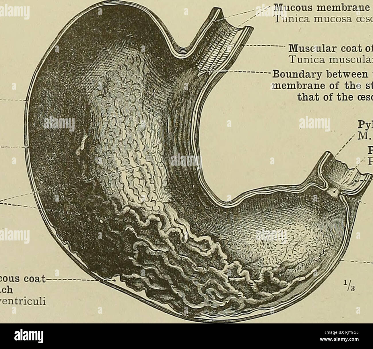
. An atlas of human anatomy for students and physicians. Anatomy. Gieat cul-de-sac, or fundus, of the stomach Fundus ventriculi Lines showing the limits of the attachment of the vis- ceral peritoneum to the wall of the stomach Body of the stomach (anterior wall) Corpus ventriculi (paries anterior) Great curvature of the stomach Cui atura ventriculi major Duodenojejunal flexure Flexura duodenojejunalis ^Inferior flexure of the duodenum- â¢Flexura duodeni inferior- ~- Jejunum Intestinum jejunum Transverse portion of the' 'Ascending portion of the duodenum i, third part) , ' duodenum (fourth part) Pars horizontalis (inferior) " ' ' Pars ascendens Inferior portion of the duodenum'* Pars inferior duodeni See Append!: : S. - See Appendix, note 6. 3 See Appendix-, note ''. Fig. 711.âVentriculus, the Stomach, moderately distended, with the Lov/est Portion OF THE CEsophagus, and the Duodenum. Seen from Before. Mucous membrane of the cesophagus Tunica mucosa cesophagi Muscular coat of the stomach â Tunica muscularis ventriculi Mucous membrane of the â stomach Tunica mucosa ventriculi Eugae of the mucous. membrane Plicae mucosa; Areolar or submucous coat of the stomach Tela submucosa ventriculi. ""- Muscular coat of the oesophagus Tunica muscularis cesophagi Boundary between the mucous membrane of the stomach and that of the oesophagus Pyloric sphincter / M. sphincter pylori Pyloric orifice Pylorus Mucous membrane of the duodenum Tunica mucosa duodeni Ridge of the pyloric ring^ Valvula pylori - Muscular coat of the stomach Tunica muscularis ventriculi â * See Appendix, note ^. Fig. 712.â-Anterior Half of the Stomach, which has been divided in Two by Incisions along the Great and Small Curvatures ; seen from the Inside. Transition of the Mucous Membrane of the CEsophagus into that of the Cardia. Pylorus, or Pyloric Orifice. ti^iCM Mucosa Ventriculi, Rug^ of the Mucous Membrane of the Stomach. Tubus digestoriusâAlimentary canal.. Please note that these i