. Biologie der Labor Maus. Mäuse als Labortiere; Mäuse; Tiere, Labor; Mäuse. 132 BIOLOGIE DER LABORMAUS fetthaltigen Zellen. Die Schleimhaut bildet niedrige längs falten. Die ureters geben Sie den dorsalen Wand des Halses der Blase zu nahe beieinander. Blase. - Die Blase wird durch Übergangsregelungen Epithel bestehend, gezeichnet, wenn die Orgel leer ist, von etwa vier bis fünf Schichten von Zellen. Die faserige lamina propria ist reich an Blutgefäßen. Die Schleimhaut ist in Breiten unregelmäßigen Falten geworfen und gelegentlich enthält eine Aggregation von Lympho-cytes. Wenn die Blase ist
1578 x 1583 px | 26,7 x 26,8 cm | 10,5 x 10,6 inches | 150dpi
Weitere Informationen:
Dieses Bild ist ein gemeinfreies Bild. Dies bedeutet, dass entweder das Urheberrecht dafür abgelaufen ist oder der Inhaber des Bildes auf sein Urheberrecht verzichtet hat. Alamy berechnet Ihnen eine Gebühr für den Zugriff auf die hochauflösende Kopie des Bildes.
Dieses Bild kann kleinere Mängel aufweisen, da es sich um ein historisches Bild oder ein Reportagebild handel
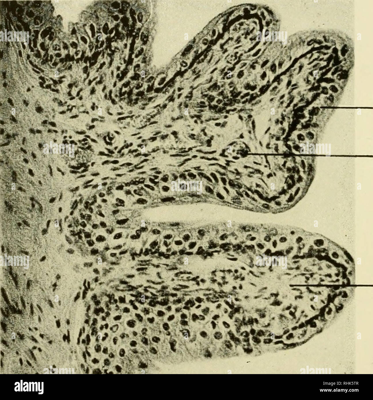
. Biology of the laboratory mouse. Mice as laboratory animals; Mice; Animals, Laboratory; Mice. 132 BIOLOGY OF THE LABORATORY MOUSE adipose cells. The mucous membrane forms low longitudinal folds. The ureters enter the dorsal wall of the neck of the bladder close to one another. Bladder.—The bladder is lined by transitional epithelium consisting, when the organ is empty, of about four to five layers of cells. The fibrous lamina propria is rich in blood vessels. The mucous membrane is thrown into wide irregular folds and occasionally contains an aggregation of lympho- cytes. When the bladder is in a distended condition the folds are absent and the epithelial lining is very thin. The smooth muscle coat consists of. Epithelium Blood vessel Lamina propria Fig. 62.—Bladder. (X200.) irregular muscle bundles of varying size, separated from each other by considerable amounts of connective tissue (Fig. 62). At the neck of the bladder the direction of the muscle bundles is circular. Female urethra.—The female urethra is a dorsoventrally slightly flat- tened tube which originates at the neck of the bladder and opens into the clitoral fossa. Near its origin the tube is lined by transitional epithelium which soon changes into stratified squamous type. The lamina propria is formed by loose connective tissue. The mucous membrane forms longitu- dinal folds. The epithelium forms invaginations which are continuous with gland tubules of the urethral glands. These glands are similar in structure to the urethral glands (of Littre) in the male. The circularly arranged smooth muscle fibers forming the outer wall are well developed. Near the. Please note that these images are extracted from scanned page images that may have been digitally enhanced for readability - coloration and appearance of these illustrations may not perfectly resemble the original work.. Roscoe B. Jackson Memorial Laboratory; Little, Clarence C. (Clarence Cook), b. 1888; Snell, George D. (George Davis), 1903-; D